eHealth Technologies Blog: Gary Larson, Executive Vice President, General Manager, HIE Solutions at eHealth Technologies
May is Stroke Month, and every member of our team is reflecting on the important work that HIEs play in the diagnosis and treatment of stroke patients.
In the time it will take you to read this blog at least one person in the US will suffer from a stroke According to the National Stroke Association. The clock starts ticking every 40 seconds for another American, as a stroke begins to deprive the brain of oxygen. From that point on, whether they fully recover, suffer through serious long-term disabilities, or die, will generally depend on the quality and speed of care received. Diagnosing and treating a stroke patient effectively depends on a carefully choreographed sequence of activities, usually starting with a medical imaging study, and frequently requiring stroke specialists who may be many miles apart to have access to those images.
eHealth Technologies has had the privilege of working with many of the leading HIEs in the nation, providing them with the ability to share medical images with care providers wherever they may be and regardless of where the image may have been generated. As such, we are in a unique position that allows us to dramatically improve the treatment effectiveness and overall outcomes for stroke – the #5 cause of death in the US, and the #1 cause of long-term disability according to the National Stroke Association.
The following scenario demonstrates the vital role that medical image sharing plays in stroke diagnosis and treatment:
When a patient presents at a local emergency department suffering with stroke symptoms, the first order of business will be to perform a brain imaging study – frequently a CT Angiography study – and have it interpreted by a neurologist or neuro-radiologist. Imaging is the only way to determine whether the patient is suffering from an ischemic stroke (a clot that impedes blood flow) or hemorrhagic stroke (bleeding around brain tissue). Each requires an entirely different treatment protocol; and for each the key to a successful outcome is diagnosing and initiating the right treatment as quickly as possible.
Time is Brain. Some ischemic strokes, which make up 85% of the total strokes in the US, require an endovascular clot retrieval procedure that is usually only performed in a JCAHO certified Comprehensive Stroke Center. Other ischemic strokes are best treated by retaining the patient at the local facility and administering clot-busting drugs such as a Tissue Plasminogen Activator (tPA).
Time is brain is a phrase frequently heard in stroke treatment circles but is much easier said than done. Critical decisions must be made quickly and accurately. Should the patient be treated locally or transferred to a Comprehensive Stroke Center? What course of treatment will be most effective – drug therapy or endovascular clot retrieval? All of these decisions must be made carefully but at lightning speed, and all are dependent on diagnostic quality medical images being readily available to neurologists who are often located many miles away, as there may be but one or two such highly-qualified specialists available in any region or state at any given time. Furthermore, these mission critical images often reside on numerous disparate hospital networks and are stored on a wide array of PACS technologies. Patients may be unconscious and known only as “John or Jane Doe.” This forces specialists to log in to one of numerous systems to access these records; and when all else fails to drive miles and miles to the hospital in the middle of the night just to view these critical images. Ancillary data, such as prior history and whether the patient is taking blood thinner medications, can also be difficult to access yet are critical to decision making. These and many other factors present a unique set of challenges that image-enabled HIEs are nearly perfectly positioned to address.
Time is brain is one of our key motivations for including Emergent Imaging Workflow in eHealth Connect® Image Exchange. With Emergent Imaging Workflow enabled for an HIE, a neurologist can access the imaging studies for every current stroke patient at all connected facilities across the community served by the HIE – all in one place and with just a few clicks. Regardless of the time of day or night, whether these highly-trained specialists are at home, in their home hospital, or travelling overseas, stroke imaging studies are accessible within minutes of being generated. This helps to assure that the most critical care decisions in stroke diagnosis and treatment are made as quickly and accurately as possible.
With Emergent Imaging Workflow, “door-to-procedure” time can be reduced significantly, minimizing the time between when a patient first presents with stroke symptoms and the optimal course of treatment being administered. Even a few minutes can mean the difference between living a life with permanent brain damage, paralysis—or even death—and a full life unimpaired by the ravages of stroke.
Because, after all, time is brain.
This month – take a few minutes to understand risk factors that you can control:
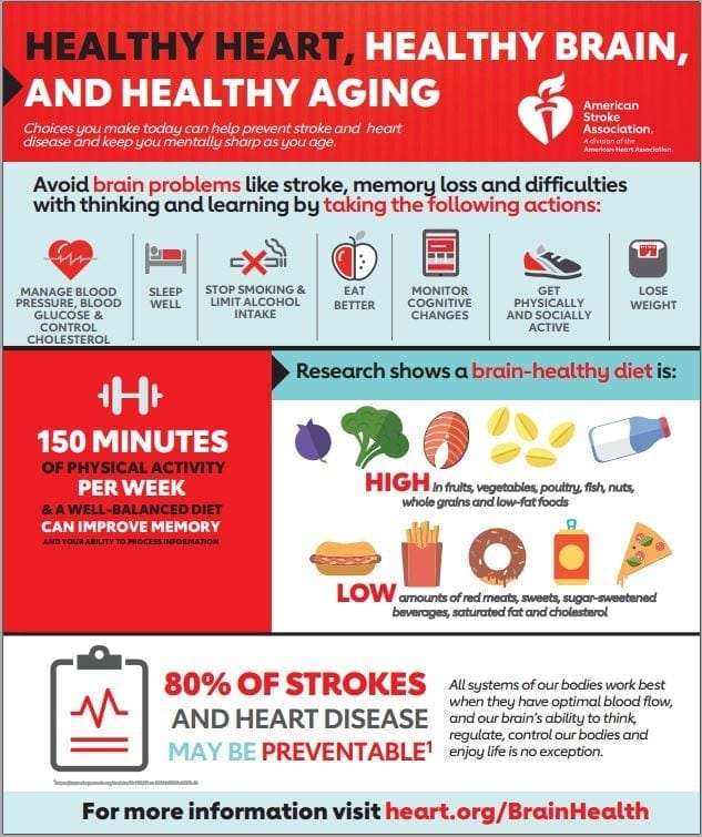
Or – take the “Stroke Risk Quiz” from the American Stroke Association.